
Lesson visuals · Diagrams
Pictures that teach
Seven original diagrams drawn for the lessons — pressure injuries, burns, injection sites, ECG and fetal circulation. Small files, crisp text, made to be read on a phone.
Skin integrity
Pressure injury — the stages
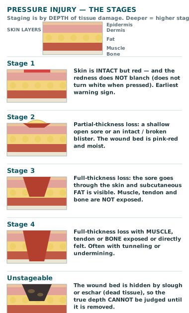
Stage by depth, not by size. A small sore can be Stage 4 if it reaches bone — and a wound covered by eschar cannot be staged at all until the dead tissue is removed.
Burns
Burns — depth of the burn
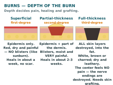
Depth decides the story. Superficial burns hurt and heal. Full-thickness burns destroy the nerve endings, so the center may not hurt at all — that silence is a danger sign, not a comfort.
Burns
Burns — rule of nines
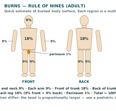
The fastest surface-area estimate in the burn unit. Count in nines — and remember this is the adult chart. Children carry proportionally more of their surface in the head.
Medication administration
IM injection sites
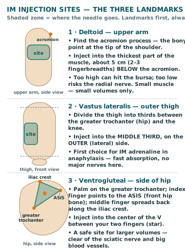
Landmarks first, needle second. Every safe IM site is found by bone landmarks — acromion, greater trochanter, iliac crest — never by guessing a spot on the skin.
Cardiac · ECG
ECG — one heartbeat, labeled
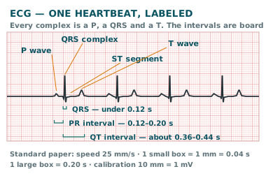
One beat, three waves, three intervals. If you can measure the PR and the QRS on a single complex, you can read any strip the exam puts in front of you.
Cardiac · ECG
ECG — the board rhythm patterns
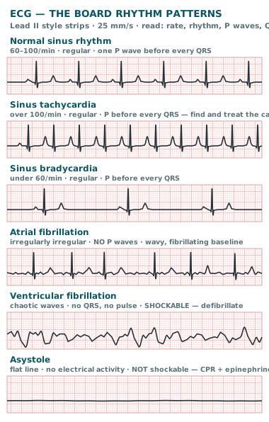
Read every strip the same way: rate, rhythm, P waves, QRS. Two of these six are act-first emergencies — VF gets a shock; asystole gets CPR and epinephrine, never a shock.
Maternal & child
Fetal circulation — the three shunts
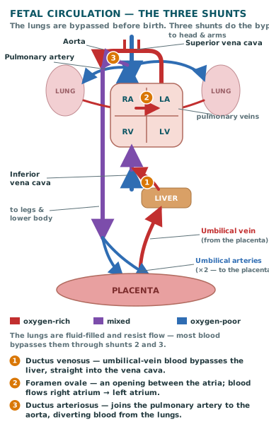
Before birth, the placenta is the lung. Three shunts route blood around the fluid-filled lungs — and all three are supposed to close once the baby takes its first breaths.